Understanding the Diagnosis
Let’s be honest – most of us have never really thought about the brain and spinal cord, except for the fact that we have one and we need to protect it. When your child or loved one is diagnosed with a neurological condition that may require neurosurgical intervention, it is helpful to understand the structure and functioning of the brain and spinal cord.
In this section we have included an overview of the anatomy of the brain and spinal cord, along with a few diagrams to help you see what it looks like. A glossary of terms can be found here.
The human brain
The Brain
The brain sits inside the skull and is surrounded by a watery fluid called cerebral spinal fluid (CSF) that provides nutrients to the brain and cushioning for protection. There are two sides to the brain: the left hemisphere and the right hemisphere. The hemispheres are responsible for different things.
Corpus callosum is the part of the brain that allows the right and left hemisphere to communicate with each other.
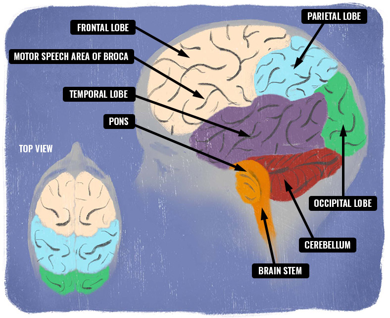
The Cerebrum
The cerebrum is the largest part of the brain and has two hemispheres (left and right). The cerebrum is extremely important as it interprets sensory input from all body parts and controls body movements. The cerebrum is responsible for movement, learning, emotions, reasoning and memory. There are four lobes that make up the cerebrum: frontal, temporal, parietal and occipital lobes. It is important to note that the movement on one side of the body is controlled by the opposite side of brain. (For example, your right arm and right leg movement is controlled by left side of your brain.)
The Four Lobes of the Brain
The frontal lobe is the lobe of the brain that helps with problem solving, judgment and decision making. This is where our personality is formed. A small part of the frontal lobe helps with speech and controls movement. The frontal lobe is located behind the forehead.
The parietal lobes help us understand and interpret information from our senses of taste, temperature, pain and touch. This area also works with handwriting, math skills, language and body position. The parietal lobe is located behind the frontal lobe.
The temporal lobes are located in the temple area. This part of the brain helps us understand what we are hearing by processing information for speech
and language.
The occipital lobes, which are located at the back of your head, deal with vision. The optic nerve carries information to the occipital lobe of the brain where it can make sense of it. The right optic nerve goes to the left occipital lobe and the left optic nerve goes to the right occipital lobe in a criss-cross.
There are special doctors at the Stollery called neuro-ophthalmologists or ophthalmologists who can assess the health of the eyes and the optic nerves. If you are scheduled for an appointment, prepare to be there for a while. The detailed examination can be lengthy, so bring extra toys and supplies.
It should not be pushed back. Protect your frontal lobe!
OTHER PARTS Of the brain
The cerebellum is at the base and the back of the brain. It is responsible for coordination, balance and motor functions such as limb and trunk movement, eye movement and speech. The cerebellum also has input into behavioural portions of the brain.
The hypothalamus is located deep within the brain and is responsible for hunger, thirst and body temperature. The hypothalamus also drives the pituitary gland to release hormones to keep the endocrine system working correctly.
The amygdala is grey matter in the medial temporal lobe and is important for emotional responses. It is part of the limbic system.
The hippocampus is important for long-term memory. It is also part of the limbic system like the amygdala and is often targeted when treating seizures surgically.
The thalamus is a deep area of the brain that relays both sensory and motor activity.
The Brain Stem
- Midbrain: This part of the brainstem is important for some eye movements and houses the wires that control movement as they travel from the brain to the spinal cord.
- Pons: This is the largest part of the brainstem and controls some eye and facial movements and sensation. It also has the very important job of relaying information from one part of the brain to other parts.
- Medulla oblongata: This is the part of the brainstem that is closest to the spinal cord and controls breathing, swallowing and heart rate. We don’t have to think about our heart beating or our lungs working because the medulla oblongata controls these involuntary functions.