Arteriovenous Malformations
Arteriovenous malformations (AVMs) are abnormal connections between the veins and arteries. This tangling of blood vessels can occur anywhere but it is most common in the brain and spine. AVMs vary in size and location in the brain.
Most individuals with an AVM do not have any symptoms until the vessels rupture and bleed into the brain. AVMs are often detected when people have a CT or MRI scan for another issue.
Symptoms of AVMs may be seizures, headaches, ear noise/buzzing, progressive weakness or numbness. More serious symptoms may require immediate medical attention, such as severe headaches, vision issues, balance issues, speech problems or weakness/numbness based on the location of the AVM.
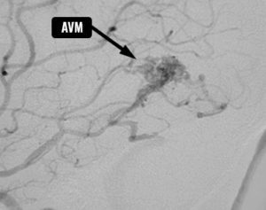
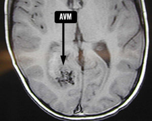
Arteriovenous malformations seen on a cerebral angiogram and MRI
Complications of AVM
- Brain hemorrhage or bleeding in the brain
- Reduced oxygen to the brain caused by the altered flow of blood in this tangled area of vessels
- Weakness in the blood vessel wall may lead to the formation of aneurysms in the veins that supply blood to the AVM
Diagnosing AVM
Any patient suspected of having an AVM will undergo a thorough history and physical exam by the medical team.
A CT scan or MRI of the brain will often be done to confirm the presence of an AVM.
Cerebral angiograms can show the location and features of the AVM. The team threads a catheter into a patient’s artery up to the site through the groin and injects a specialized dye to map out the vessels.
Treatment
A neurosurgeon will decide how to treat the AVM depending on the location and size of the abnormal blood vessels.
If surgery isn’t needed, the doctor may order medications to help manage symptoms.
- Surgical removalof the AVM might be in order if it’s small and not deep within brain tissue. The neurosurgeon would remove a part of the skull to gain access to the brain, then use special clips to seal off the AVM and remove it. The surgeon would reattach the skull and close the scalp incision.
- Endovascular embolizationis a procedure in which the doctor uses advanced x-ray machinery to thread a catheter through the leg artery up to the brain. The doctor places the catheter in one of the arteries that feeds the AVM and he injects a glue-like substance to reduce the blood flow to the AVM. Endovascular embolization occasionally is used as a stand-alone treatment but most often is coupled with surgery or stereotactic radiosurgery.
- Stereotactic radiosurgery is a procedure that uses focused radiation to scar the area and shrink the AVM. Generally AVMs smaller than three centimetres are suitable for this technology and it takes a minimum of two to three years to know if the treatment has worked.
Reference:
mayoclinic.org/diseases-conditions/brain-avm/basics/definition/con-20034230 Reviewed April 2, 2014; kidshealth.org/en/parents/arteriovenous-malformations.html