CSF Shunts
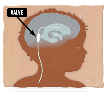
A shunt is a tube, placed in by a neurosurgeon, that allows the fluid to flow through it. A valve regulates the flow of the fluid. Most shunts are used to divert cerebrospinal fluid from one location to another. Mostly, shunts have three pieces: a proximal catheter, a valve mechanism (the bump under the skin on the head that may be located at the top or back of the head) and a distal catheter. The neurosurgeons at the Stollery are very experienced with shunts. It is important to avoid constipation with shunts, as it increases the pressure in the abdomen, making it difficult to drain CSF from the brain.
Shunt types
- Cerebral shunt: Some children have increased fluid around their brain
or conditions that cause increased intracranial pressure. In these
children, a one-way valve is used to drain excess fluid from the brain and carry it to other parts of the body. A one-way valve means the fluid can only travel one way: out of the brain. This valve sits outside the skull, but beneath the skin. - Lumbar-peritoneal shunt: This is a tube or shunt with a one-way valve that is used to drain the spinal cerebrospinal fluid and transports it to the abdomen. This shunt is usually inserted in the lower back (lumbar area). It then runs beneath the skin to the abdomen, and eventually to the abdomen (belly) where it gets absorbed.
Shunt Type
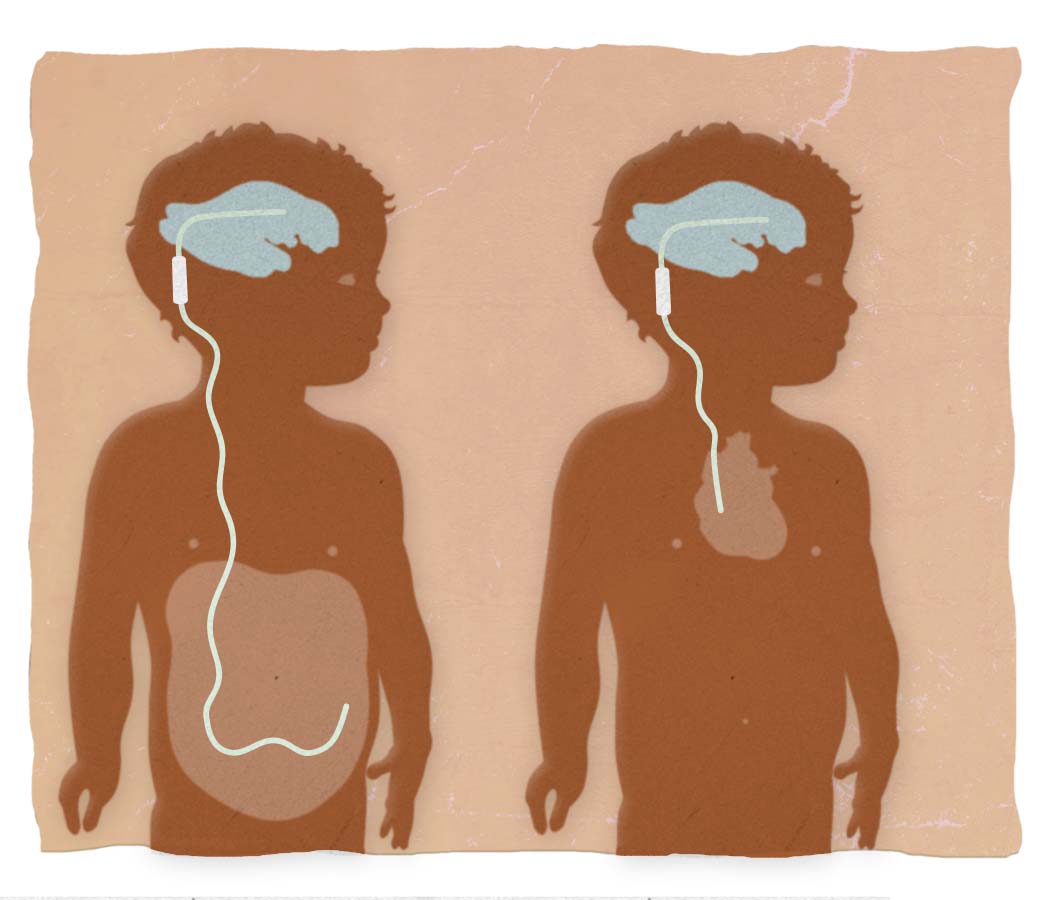
- Ventriculo-peritoneal (VP) shunt: Inserted into the ventricle of the brain and drains into the peritoneal cavity (abdomen)
- Ventriculo-atrial (VA) shunt: Inserted into the ventricle of the brain and drains into the right atrium of the heart
- Ventriculo-pleural (VPL) shunt: Inserted into the ventricle of the brain and drains into the pleural space (chest)
- Lumbar-peritoneal (LP) shunt: Inserted in lower back (lumbar) spinal subarachnoid space and drains into the peritoneal cavity
GOOD TO KNOW: If the shunt is placed in the lumbar area of the spine, the bump from the valve will be on the back next to the spine.
If your child has a programmable shunt, a neurosurgeon may need to reprogram it after the MRI is complete. An MRI machine uses a magnetic field, which may change the setting of your child’s shunt. NEw programmable valves are being developed so that reprogramming is not necessary after an MRI scan.
Children with programmable shunts should stay away from strong magnets. Toys like Thomas the Train have very weak magnets and are okay, but there are many strong magnetic devices on the market to avoid.
Tests and scans used for shunts
- Shunt series: These are x-rays used to examine the tubing. They help identify if there is a disconnection or a breakage.
- MRI scans: There are two types of MRI scans. First, there is a fast head MRI (FISP), which takes about 30 seconds, and your child will be awake. Parents can accompany child if required. This scan is used to determine if there is a change in the size of the ventricles. Second, a full MRI is a more detailed MRI that can take 30-60 minutes and examines the CSF flow as well as the brain itself. Sedation is frequently required for small children.
- CT scan: a quick scan, useful in looking at the tubing of the shunt in the brain as well as in identifying any bleeding or blood in the ventricle.
What are they looking at?
Here is the picture of a normal brain MRI.
In the middle of the brain you’ll see a darkened thin shape that resembles bat wings. This is the ventricle system of the brain.
Now, look at the views below that show hydrocephalus on a CT, MRI and fast head MRI (FISP scan or fast head). In hydrocephalus, you can see how the ventricles (bat wings) are much bigger than in a normal brain, and now resemble a butterfly.
Doctors will compare older and new CT or MRI films to assess if the ventricles are remaining stable in size or if they have changed. This information, along with your child’s symptoms, allows the neurosurgical team to know if there is a problem with the functioning of the shunt.


GOOD TO KNOW: Neither kind of MRI has radiation.
Shunt tap
Most shunt valves have a small dome or bulb reservoir (this is the bump you see under the skin) designed to sample CSF by inserting a small needle under sterile procedure by a neurosurgeon or NP. This procedure is done to relieve pressure in urgent situations or to test for infection.
The Stollery’s neurosurgery team identified some signs and symptoms of shunt malfunction or infection to watch for:
- Signs and symptoms for infants
- Enlargement of baby’s head
- The soft spot at top of head is full and bulging
- Prominent scalp veins
- Swelling around shunt
- Vomiting
- Irritability
- Sleepiness
- Not eating or drinking
- Redness along shunt tract
- Downward gazing of the eyes
- Fever
- Seizures
- Signs and symptoms for toddlers
- Head enlargement
- Vomiting
- Headache
- Irritability
- Sleepiness
- Loss of previous ability
- Swelling on shunt tract
- Fever
- Seizures
- Signs and symptoms for children and adolescents
- Vomiting
- Headache
- Vision Problems
- Irritability and/or fatigue
- Personality changes
- Loss of coordination or balance
- Swelling at shunt
- Difficulty at waking up or staying awake
- Decline in academic performance
- Seizures
- Fever
- Redness along shunt
You may be hesitant to seek medical attention out of fear that you are overreacting to your child’s symptoms. Children with shunts can get sick very quickly, so it’s better to be safe than sorry. This may result in some ER visits where they are seen, assessed and sent home with the assurance that everything is fine. By having your child assessed properly when symptomatic, when your child’s shunt fails they will receive the life-saving, prompt medical attention they require.
TIP: A simple way to prevent shunt infections is to cut your child’s fingernails! Children will often scratch at their shunts and, if the scratch is deep enough, their shunt can get infected.
Shunt revision
When a shunt needs to be replaced it is called a shunt revision. There are many reasons a shunt needs to be revised:
- Infection
- A blockage in the shunt system
- The shunt is over or under draining for the needs of the child
- The catheter is no longer in an ideal position
- Tubing is broken
This is an example of a ventricular catheter (shunt) that has failed due to the in-growth of the choroid plexus, which is like the “seaweed” of the brain that creates the cerebrospinal fluid. It can plug the catheter and prevent CSF from entering and draining.
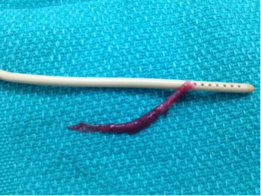
Failed ventricular catheter (shunt)
Signs and symptoms after a shunt insertion or revision
After a shunt operation, you need to watch for infection or shunt malfunction. Seek medical help if you notice any of the following:
- Fever (greater than 38 C)
- Redness, swelling or pustules along shunt
- Intense abdominal (tummy) pain
- Headache after first seven to 10 days
- Unusually tired and/or irritable
- Vision problems
- Fluid collecting or leaking along shunt tract
- Bulging at the valve on the head
- Seizures
Use a chart like this one to record information about your child’s shunt: the date of surgery or change, the kind of shunt it is, and the setting on the shunt.
Reference:
Toporck, C. & Robinson, K. (1999). Hydrocephalus: A Guide for Parents, Families and Friends. O’Reilly and Associates Inc.